Medical Imaging Workflows for Clinical AI
Built for clinical AI startups and researchers. Generate synthetic data, support segmentation-aware model training, and build evaluation workflows with reliable ground truth when needed.
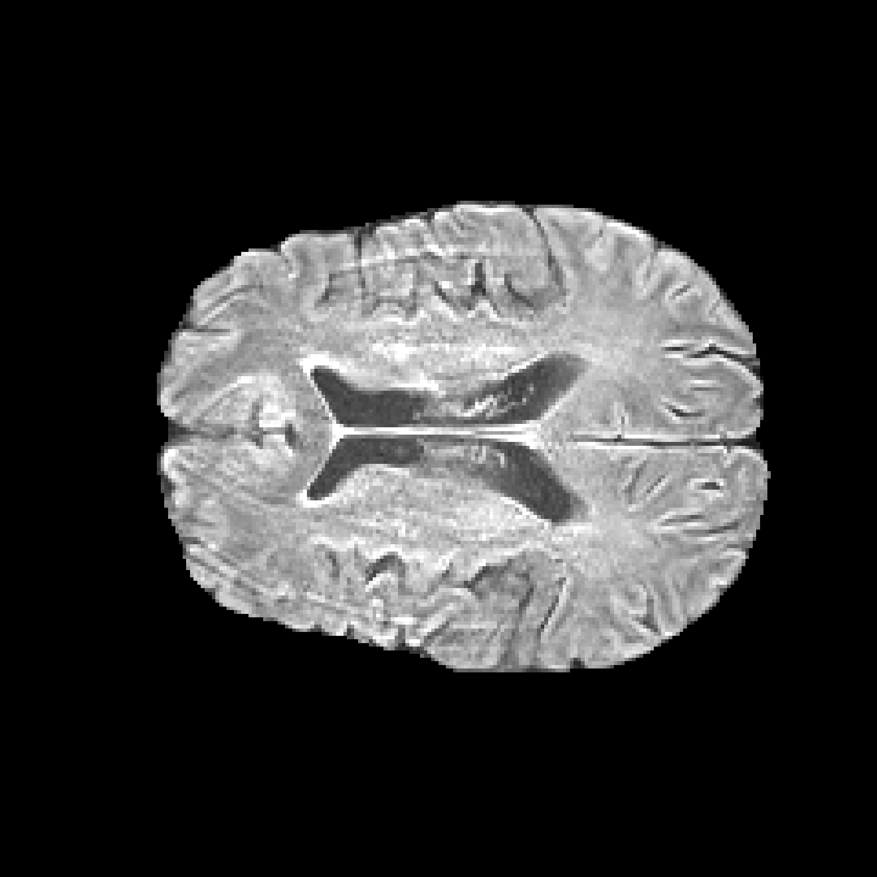
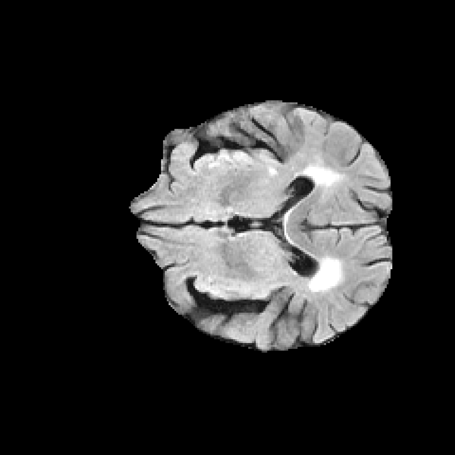
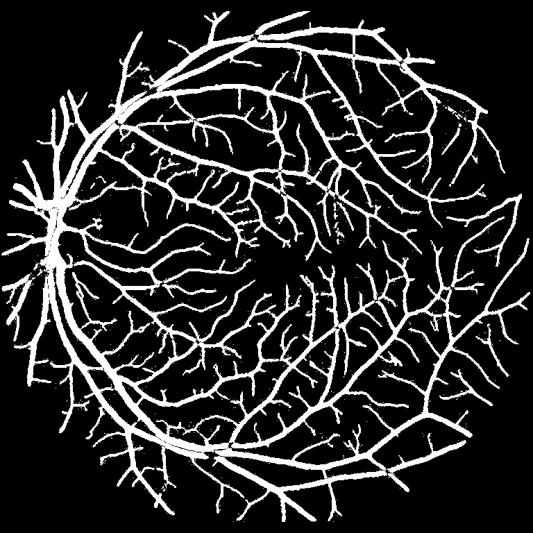
Sample Output
The Problem
Medical AI Development Is Stuck on Data
Current medical AI development is constrained by limited access to high-quality clinical imaging data, especially for validation where datasets must be independent, unbiased, and acceptable to regulators.
$50k–$500k per FDA Submission
Medical AI startups spend hundreds of thousands acquiring and annotating data for a single regulatory submission.
Months of Data Collection
Obtaining high-quality, annotated clinical imaging data is slow, requiring IRB approvals and data use agreements.
Privacy & Compliance Barriers
Medical data is highly sensitive, requiring HIPAA and GDPR compliance, making it difficult to access and use.
Rare Pathologies Are Scarce
Edge cases and rare conditions are underrepresented in clinical datasets, limiting model robustness.
Why Physics-First
The Physics-First Difference
Unlike generative AI, every synthetic image is deterministically rendered from anatomical and optical models. No mode collapse. No artifacts. Full traceability.
Parameters define disease state and vascular structure before image formation. Every pixel is traceable to a physiological model.
Ray-tracing and fluid dynamics simulate the actual imaging device optics. No shortcuts, no hallucinations.
Traceable ground truth accepted as evidence for FDA MDDT pathways. Built for the scrutiny of clinical deployment.
How It Works
One API Call. Clinically Accurate Output.
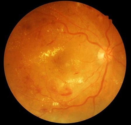
Choose your imaging modality, send anatomy parameters, and receive publication-ready synthetic images with complete ground-truth maps.
Send anatomy parameters, receive publication-ready synthetic fundus images with complete ground-truth segmentation maps.
# Python SDK
from medsim import Client

client = Client("sk_live_...")

result = client.fundus.generate(
  pathology="diabetic_retinopathy",
  severity="moderate",
  resolution=2048,
  demographics={
    "age": 62,
    "ethnicity": "south_asian"
  }
){
"id": "img_8f3k2m9x",
"status": "complete",
"image_url": "https://cdn...",
"ground_truth": {
"vessel_map": "https://cdn...",
"lesion_mask": "https://cdn..."
},
"render_time_ms": 1247
}
Use Cases
Built for Clinical AI Startups & Researchers
From academic research through clinical deployment and regulatory submission, MedSim API integrates into your existing workflows.
Accelerate Medical Research Without Data Bottlenecks
Whether you're studying Alzheimer's biomarkers in retinal imaging or brain MRI, characterizing rare pathologies across modalities, or analyzing retinal layer changes in OCT — MedSim API gives researchers instant access to controlled, labeled synthetic data. No IRB approvals. No months-long data agreements.
- Generate disease-specific datasets for any research hypothesis
- Study progression across severity levels with controlled parameters
- Perfect ground truth for quantitative analysis and paper-ready figures
- Academic pricing available for .edu-affiliated researchers
Segmentation Samples
Original Images and Vessel Masks
Alongside synthetic data generation, we use our own AI models to generate segmentation outputs. These examples show original fundus images paired with vessel masks for training, evaluation, and visual quality review.
Left Eye Vessel Segmentation
Drag across the image or use the slider bar to compare the original image and vessel mask.
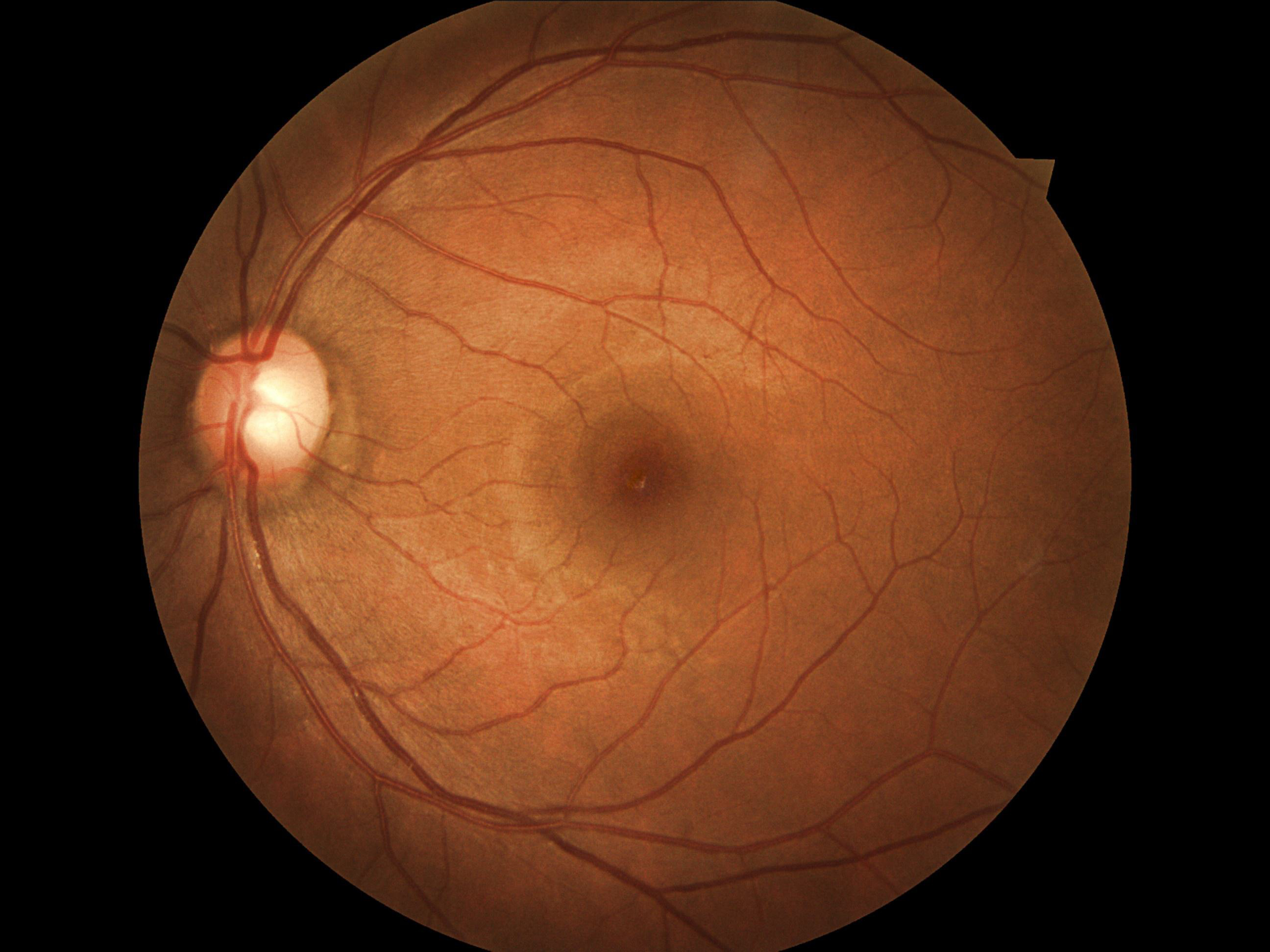
Original left-eye fundus image with a corresponding vessel mask highlighting the retinal vasculature.
Browse real segmentation samples
Explore several image-and-mask pairs to see how vessel segmentations align with the underlying retinal structures across different samples.
Validation
Experimentally Validated
We've proven that our physics-first approach produces synthetic data suitable for AI training, validation, and regulatory submission.
Proven Performance
Our synthetic fundus images trained a vessel segmentation model that achieved performance parity with real-world data on DRIVE and STARE clinical datasets.
Zero Labeling Cost
Perfect ground truth segmentations, lesion boundaries, and quantitative biomarkers are generated automatically—no manual annotation required.
Scalable & Reproducible
Generate thousands of high-fidelity images with deterministic, version-controlled parameters for reproducible benchmarks.
Pricing
Simple, Transparent Pricing
Pay per image with no upfront commitment. Academic researchers with a .edu email get a 40% discount — because breakthroughs shouldn't be gated by budget.
For academic researchers and students with a .edu email. Full API access at a discounted rate.
~$3 per 100 images
- All imaging modalities (Fundus, MRI, OCT)
- Full ground-truth segmentation maps
- Up to 10,000 images / month
- Python SDK & REST API access
- Priority email support
- Dataset export in standard formats
For clinical AI startups building and validating models for production deployment.
~$5 per 100 images
- All imaging modalities (Fundus, MRI, OCT)
- Full ground-truth segmentation maps
- Up to 100,000 images / month
- Python SDK & REST API access
- Priority support with SLA
- Batch generation & webhooks
- Deterministic versioned datasets
For organizations needing custom modalities, volume pricing, and regulatory support.
Tailored to your needs
- Everything in Startup, plus:
- Unlimited image generation
- Custom modality development
- On-premise deployment option
- Dedicated account manager
- Regulatory documentation support
- Custom SLA & compliance review
Need high-volume batch generation?
All plans support batch API calls. Generate up to 1,000 images per request with automatic queuing and webhook notifications. Volume discounts available for batches over 50,000 images.
FAQ